Posts

CHRONIC BRONCHITIS

Image
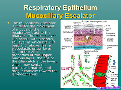
  Chronic Bronchitis : Inflammation and swelling of the lining of the airways, leading to narrowing and obstruction generally resulting in daily cough. Chronic Bronchitis - Muco-ciliary escalator  In heavy smoker ➝ Tar ↓ Ciliary damage ↓ Failure of muco - ciliary escalator ↓ Stasis of secretion ↓ infection ↓ Pus ↓ Occlusion of respiratory bronchiole Leading to underventilation of alveoli ↓ Very severe hypoxia ⟶ Central Cyanosis (↓↓ PO2) Increased pCO2 ➝ Narcosis , sleeping period increase , weight gain - 'BLUE BLOATERS' Clinical Feature : Wheel chair bound patients Type 2 Respiratory failure Respiratory acidosis Bronchorrhea central cyanosis On Examination : Increased Respiratory Rate Accessory muscles overactivity , subcostal recession Pursed lips, Central Cyanosis(blue bloater) Halitosis Clubbing absent Both inspiratory and expiratory Ronchi Hemoptysis is absent Work up : Spirometry Chest X ray - increased Broncho vascular markings(dirty lungs) IOC - HRCT ABG - Type 2 Respira

EMPHYSEMA

Image
 Emphysema is a type of  COPD  (chronic obstructive pulmonary disease). Emphysema is defined as enlargement of the air spaces distal to the terminal bronchioles, with destruction of their walls of the alveoli. Blebs in respiratory airways  Type 1 respiratory failure respiratory alkalosis Air trapping on exhalation and amount of air going out is lesser ↓ So FEV1 less than normal  ↓ On giving Salbutamol, no change in FEV1 So it is a Non Reversible Air flow obstruction On examination : Breathlessness Exercise intolerance Tachypnoea Accessory muscle working excessively Barrel shaped chest ( anterioposterior diameter > Transverse diameter) Liver palpable (due to hyperinflation of lungs) work up : Spirometry HRCT (investigation of choice) - Blebs ABG - Type 1 Respiratory failure (Respiratory Alkalosis) DLco - Diffusion capacity of lung for carbon monoxide - decreased chest x-ray - Flattening of diaphragm - Tubular heart - Lungs appear more black than normal - Vascular markings present Tre

BRONCHIAL ASTHMA

Image
Main diagnostic criteria for bronchial asthma is reversibility of airflow obstruction which is demonstrated by spirometry.                               SPIROMETER : Patient is made to perform forceful inspiration and expiration and flow volume curves are obtained Baseline curve for inspiration part remain same but expiration part patient is not able to push out the air as asthma is disease of expiratory. so, marked concavity in expiration part is present Now salbutamol is given and test is repeated in 10 - 15 min, concavity will reduce significantly (see above) FEV1 decreased ⟶⟶ after S.A.B.A (short acting beta2 agonist) ➝➝ FEV1 normal Methacholine provocation test : It is of historical importance. not done now as may lead to imminent respiratory failure. Eucapnic hyperventilation test : Person is made to breathe in cold air. asthmatics has bronchial hyper reactivity to cold air and FEV1 will fall DIAGNOSTIC CRITERIA : Reversibility (Absolute criteria): asthma is reversible airflow ob

COMMUNITY ACQUIRED PNEUMONIA

Image
Acute infection of pulmonary parenchyma Causative Organism : Streptococcus pneumoniae (pneumococci) - most common Haemophilus influenzae - 2nd most common Mycoplasma pneumonia Chlamydia pneumonia Rare : Legionella Staphylococcus aureus Gram negative organism ( klebsiella , pseudomonas) virus (adenovirus , meta pneumo virus) In India : - 20% cases of CAP - Tuberculosis - 40% cases of CAP - Not established PATHOLOGY : Initially there is a inhalational of bacteria into the lungs results in phase of Edema  ↓  proteinaceous exudates developed in the alveoli (so there can be a crepitation, crackles of  bronchial breathing in the patient and subsequently patients enter into the following phases) Red hepatization - RBC + Neutrophils (3-4 days) Grey hepatization - RBC lysis + Neutrophils (it signifies successful)(5 -7 days) Resolution - here Macrophages predominantly present because these cells restore the normal tissue Typical / lobar pneumonia : Lobar consolidation causative organism ; - Pneu

BRONCHIECTASIS

Image
Bronchiectasis : Destruction / Dilatation of large airways - airways are filled with pus results in the features of bronchorrhea HISTOPATHOLOGICAL  EXAMINATION: CYLINDRICAL / TUBULAR - MOST COMMON subtype - Represents airways are dilated and thick walled which filled with pus by the dangerous organisms like pseudomonas. this organism later will form the biofilm in the lungs which results in antibiotic resistant. expansion of pus filled cavity will erode the blood vessels of lungs which results is massive hemoptysis and death of the patient. Cause of  death - Massive hemoptysis Causes : A) FOCAL - Obstruction which subsequently cause infection in the airway and results in pus. following condition can cause focal bronchiectasis - Foreign body aspiration (children) - Pulmonary hamartoma (adults) - Bronchial carcinoid (adults) note: Benign tumor in adults like pulmonary hamartoma and carcinoid tumors which encroached the bronchus and result in obstruction of airways and causes atelectasis.

CYSTIC FIBROSIS

Image
 It is also known as MUCOVISCIDOSIS (because the secretions in body especially the one in relation with the pancreas, are very thick and contribute to various manifestation) It is also known as EXOCRINOPATHY (because this condition is having the propensity to involve the exocrine gland) PATHOGENESIS : AUTOSOMAL RECESSIVE Defect chromosome 7 It is caused by the presence of mutations in both copies of the gene for the cystic fibrosis transmembrane conductance receptor (CFTR) protein.  In cystic fibrosis - Phenyl alanine present at 508th position is deleted so, most common mutation if F508 mutation CFTR protein malfunction leads multiple manifestations chloride channel is defective, as a result of which chloride/bicarbonate/water transport doesn't take place across it and due to this periciliary dehydration occurs and mucus becomes highly viscid Important organs involved in this conditions: Lungs GIT Pancreas - results in type 2 diabetic mellitus in future Liver - biliary stasis can c

HYPOTHYROIDISM

Image
 Hypothyroidism(underactive thyroid) is a condition in which thyroid gland doesn't produce enough thyroid hormone PRIMARY HYPOTHYROIDISM Thyroid gland is involved T4 DECREASED , T3 DECREASED , TSH  INCREASED(T4↓,T3↓,TSH↑) CAUSES : INDIA most common cause HASHIMOTO THYROIDITIS ,  GLOBALLY - ENDEMIC GOITER ENDEMIC CRETINISM - Body with hypothyroidism since birth Food goitrogen - cabbage ,  Drug induced hypothyroidism : Amiodarone SECONDARY HYPOTHYROIDISM Pituitary gland is involved T4 DECREASED , T3 DECREASED , TSH DECREASED(T4↓,T3↓,TSH↓) CAUSES : SHEEHAN SYNDROME (Pituitary infarction) Non  obstetric cause of pituitary damage called as simmond's disease - occurs due to AVM rupture ,HTN , Trauma , sickle cell anemia Cranial radiation/sarcoidosis Empty sella syndrome / CSF leak syndrome  CLINICAL FEATURES : Cold intolerance Myxedema (alopecia , puffy face , Hoarseness voice , thick coarse skin , constipation) Menorrhagia and infertility Headache Galactorrhea (TRH stimulate anterio