Posts

Showing posts with the label #TREATMENT

ACROMEGALY

Image
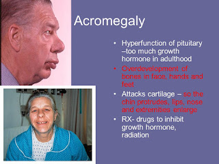
ACROMEGALY It is a disorder that results from excess growth hormone (GH) after the growth plates have closed (excess GH after puberty) It is a diseases of anterior pituitary Somatotrophs are abundant cells in pituitary somatotroph adenoma is most common cause of developing Acromegaly Mammosomatotroph adenoma growth hormone and prolactin increases clinical features : Galactorrhea Leading cause of extra pituitary source of excess growth hormone → pancreatic adenoma (islet cells tumor) Leading cause of excessive GHRH production → CARCINOID TUMOR CLINICAL FEATURES : ACRAL ENLARGEMENT : Increase in size of hand/feet Increase in finger thickness leading to spade like hands Lower Jaw prominence called as Prognathism Frontal Bossing Heel pad thickness >21 mm → used for monitoring growth in these patients Coarse facies / deep hollow voice ,Hyperhidrosis(excessive sweating) , sebum increase (increase incidence of acne) , ac...

SHEEHAN SYNDROME

Image
SHEEHAN SYNDROME It is also known as SIMMOND'S SYNDROME / POSTPARTUM HYPOPITUITARISM / POSTPARTUM PITUITARY GLAND NECROSIS Postpartum hemorrhage ↓ causes pituitary damage ↓ results in secondary addison's disease Results in decrease ACTH ↓ decrease cortisol ↓ Hypoglycemia Blood pressure is normal as aldosterone is not affected No hyperpigmentation as ACTH levels are low TREATMENT: DEXAMETHASONE

WATERHOUSE-FRIDERICHSEN SYNDROME

Image
WATERHOUSE-FRIDERICHSEN SYNDROME It is also known as HEMORRHAGIC ADRENALITIS / FULMINANT MENINGOCOCCEMIA It is defined as adrenal gland failure due to bleeding into the adrenal glands,commonly caused by severe bacterial. It is caused by Neisseria Meningitidis / Pseudomonas Leads to Sepsis DIC (Disseminated intravascular coagulation) → adrenal hemorrhage → leading to Addisonian crisis TREATMENT : I.V  Hydrocortisone + Antibiotics + Fluid replacement

ADDISON'S DISEASE

Image
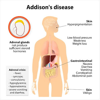
ADDISON'S DISEASE It is also known as PRIMARY ADRENAL INSUFFICIENCY / HYPOCORTISOLISM It is a long term endocrine disorder in which the adrenal glands do not produce enough steroid hormone(cortisol and aldosterone) Leading cause  → Autoimmune destruction most common cause in india → Tuberculosis of adrenals CLINICAL FEATURES : Aldosterone decrease resulting in : Salt wasting , craving for salty foods polyuria (urine output >3L/day) postural hypotension hyperkalemia metabolic acidosis Cortisol decrease resulting in : cortisol deficiency results in decreased blood sugar → palpitations,tremors,diaphoresis,RAGE attack,emotional fragility ACTH increased : Have partial melanocyte stimulating hormone like action resulting in Hyperpigmentation at palmar / sole creases scars / areola / axilla / cubital fossa groin area oral mucosa nails DHEAS (Dehydroepiandrosterone sulfate) decreased resulting in : decrease libido er...

LIDDLE SYNDROME

Image
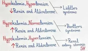
LIDDLE SYNDROME It is a AUTOSOMAL DOMINANT disorder It involving increased activity of the epithelial sodium channel(ENaC) Causes hypertension → negative inhibition of RAAS( renin angiotensin aldosterone system) system → renin decreased → aldosterone decreased  By this feedback mechanism aldosterone levels are decreased but hypertension occur due to excess salt and water in the body secondary to excessive activity of epithelial sodium channel Imaging : shows normal adrenal gland DOC : AMILORIDE (inhibit function of epithelial sodium channel

HYPERALDOSTERONISM

Image
HYPERALDOSTERONISM It is a medical condition wherein too much aldosterone is produced by adrenal gland Hyperaldosteronism can be "primary" and "secondary" Primary hyperaldosteronism Excess production of aldosterone due to Bilateral adrenal hyperplasia (most common cause) Adrenal adenoma(tumor in the first layer of cortex)→ CONN'S SYNDROME   Features: increase aldosterone cause more stimulation of epithelium sodium channel (E NaC) resulting in Increase sodium and water absorption resulting in expansion of plasma volume contralateral urinary loss of potassium and hydrogen clinical features: Headache due to hypertension Urinary loss of potassium(hypokalemia) resulting in muscle cramps(weakness) metabolic alkalosis due to loss of hydrogen ions gain of water in the body → expansion of plasma volume which increase venous return → ANF release(atrial natriuretic factor)→ natriuresis(loss of water) so, gain of water = loss of water(...

HEPATITIS D (Delta)

Image
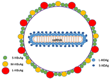
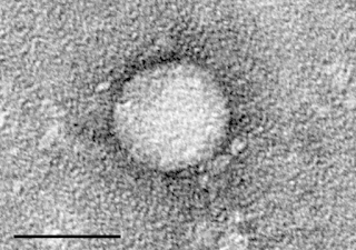
HEPATITIS D It is a disease caused by the HEPATITIS DELTA VIRUS (HDV) A small spherical enveloped virusoid Weak virus → for eradication 100% coverage of population with HBV vaccine should be done Route of spread - parenteral Transmission of HDV can occur either simultaneous infection with HBV (coinfection) or superimposed on chronic hepatitis B or hepatitis B carrier state (superinfection)

HEPATITIS C

Image
HEPATITIS C It is an infectious disease caused by the hepatitis c virus (HCV) that primarily affects the liver. most common cause of transmission is parenteral IV drug abusers(IVDA) > B T (blood transfusion) 90% cases of hepatitis C develops chronic hepatitis ↓↓↓↓ Activation of stellate cells ↓↓↓↓ cirrhosis - E.S.L.D (end stage liver disease) ↓↓↓↓ Liver transplant  (most common cause for liver transplant in world is HCV induced cirrhosis) Investigation of choice(IOC) : E.I.A (enzyme immuno assay) anti HCV antibody PCR HCV RNA : "no cut off"  for management of patient TREATMENT: Treatment based on Genotype Genotype 1: DOC - Ledipasvir Genotype 2: Sofosbuvir + Ribavirin Note: Pegylated interferon + Ribavirin (older)

HEPATITIS - B

Image
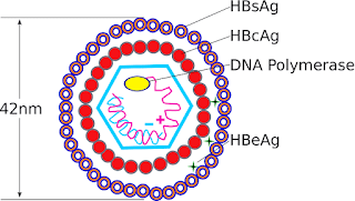
HEPATITIS - B It is a viral infection that attacks the liver and can cause both acute and chronic disease On electron microscope view virus is either spherical or tubular in shape HBsAg  -  surface antigen HBcAg  -  core antigen → found in nucleus of hepatocytes but never found in blood however antibody to it is seen in blood HBV DNA HBeAg  -  marker for replication/infectivity of virus Pre - core mutant = HBeAg absent - More severe - Replication not detected routinely ROUTE OF TRANSMISSION: Most commonly - percutaneous route : sexual transmission Blood transfusion Vertical transmission - mother to baby Accidental needle stick injury SERUM MARKERS: HBsAg -  - first to appear 1 - 12 weeks of infection - earliest serological evidence of hepatitis - B infection - precedes SGPT increases = 2 - 6 weeks - sequence of appearance HBsAg positive → SGPT increases → jaundice → disappear in reverse direct...

RENAL ARTERY STENOSIS

Image
RENAL ARTERY STENOSIS Also known as Reno vascular hypertension It is the narrowing of one or both of the renal arteries ETIOLOGY: most common cause - Atherosclerosis fibromuscular dysplasia (FMD) - (young females) Takayasu arteritis  - Asian population (pulseless disease) CLINICAL FEATURE: Refractory hypertension sudden rise BP in preexisting HTN patient flash pulmonary edema presence of abdominal bruit (systole + diastole) check peripheral pulses poly arteritis nodosa - not cause of renal artery stenosis but can lead to Aneurysm formation WORKUP: USG - Asymmetric kidneys size - Not responding to anti hypertensive medication Doppler - best screening test CT Angiography Magnetic Resonant Angiography (MRA) - expensive ↓↓↓  - false positive results - due to turbulent flow - Gadolinium - cause nephrogenic system fibrosis IOC - Renal angiography (invasive test)(gold standard) DTPA scan before and after giving ACE inhibitor (Capt...

BERGER'S DISEASE

Image
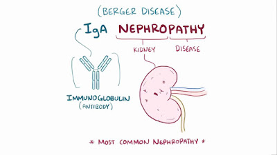
BERGER'S DISEASE It is also known as IgA Nephropathy first line of defence - IgA (Mucosal immunity) most common form of glomerulonephritis worldwide In berger's disease IgA increased with diffuse mesangial deposits - Capillaries - normal - golmerular filtration rate - normal - blood pressure - normal - microscopic Hematuria - If patient develops URTI (Upper respiratory tract infection) : IgA levels rise and over next 48 hours, microscopic hematuria is replaced by macroscopic / gross hematuria O/E - BP normal, no edema is noted WORKUP: >3RBC / HPF C3 normal in most cases TREATMENT: No consensus on optimal treatment

NEPHRITIC SYNDROME

Image
NEPHRITIC SYNDROME It is a syndrome comprising signs of nephritis, which is kidney disease involving inflammation. it often occurs in the glomerulus, which is called glomerulonephritis Vasculitis - GFR decreased and renin increased leading to - Hypertension - Hematuria - Sub nephrotic proteinuria POST STREPTOCOCCAL GLOMERULONEPHRITIS (PSGN) Type III Hypersensitivity reaction Immunological manifestation seen with nephritogenic stain - streptococcus Antigen - streptococcal pyrogenic exotoxin B (SPEP) Immune complex (+) - complement mediated vasculitis PSGN - begin as impetigo or pharyngitis Hematuria cola coloured urine - smoky urine(freshly voided sample) Hypertension - sudden onset - left ventricular failure Dyspnea - pulmonary edema Renal insufficiency leading to feature of uremia WORKUP: Urine microscopic - >3 RBC/HPF (high power field) - RBC casts - Dysmorphic RBC - always indicate glomerular bleeding C3 decreased, CH50...