Posts

Showing posts from May, 2020

LIDDLE SYNDROME

Image
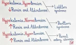
LIDDLE SYNDROME It is a AUTOSOMAL DOMINANT disorder It involving increased activity of the epithelial sodium channel(ENaC) Causes hypertension → negative inhibition of RAAS( renin angiotensin aldosterone system) system → renin decreased → aldosterone decreased  By this feedback mechanism aldosterone levels are decreased but hypertension occur due to excess salt and water in the body secondary to excessive activity of epithelial sodium channel Imaging : shows normal adrenal gland DOC : AMILORIDE (inhibit function of epithelial sodium channel

HYPERALDOSTERONISM

Image
HYPERALDOSTERONISM It is a medical condition wherein too much aldosterone is produced by adrenal gland Hyperaldosteronism can be "primary" and "secondary" Primary hyperaldosteronism Excess production of aldosterone due to Bilateral adrenal hyperplasia (most common cause) Adrenal adenoma(tumor in the first layer of cortex)→ CONN'S SYNDROME   Features: increase aldosterone cause more stimulation of epithelium sodium channel (E NaC) resulting in Increase sodium and water absorption resulting in expansion of plasma volume contralateral urinary loss of potassium and hydrogen clinical features: Headache due to hypertension Urinary loss of potassium(hypokalemia) resulting in muscle cramps(weakness) metabolic alkalosis due to loss of hydrogen ions gain of water in the body → expansion of plasma volume which increase venous return → ANF release(atrial natriuretic factor)→ natriuresis(loss of water) so, gain of water = loss of water(

HEPATITIS D (Delta)

Image
HEPATITIS D It is a disease caused by the HEPATITIS DELTA VIRUS (HDV) A small spherical enveloped virusoid Weak virus → for eradication 100% coverage of population with HBV vaccine should be done Route of spread - parenteral Transmission of HDV can occur either simultaneous infection with HBV (coinfection) or superimposed on chronic hepatitis B or hepatitis B carrier state (superinfection)

HEPATITIS C

Image
HEPATITIS C It is an infectious disease caused by the hepatitis c virus (HCV) that primarily affects the liver. most common cause of transmission is parenteral IV drug abusers(IVDA) > B T (blood transfusion) 90% cases of hepatitis C develops chronic hepatitis ↓↓↓↓ Activation of stellate cells ↓↓↓↓ cirrhosis - E.S.L.D (end stage liver disease) ↓↓↓↓ Liver transplant  (most common cause for liver transplant in world is HCV induced cirrhosis) Investigation of choice(IOC) : E.I.A (enzyme immuno assay) anti HCV antibody PCR HCV RNA : "no cut off"  for management of patient TREATMENT: Treatment based on Genotype Genotype 1: DOC - Ledipasvir Genotype 2: Sofosbuvir + Ribavirin Note: Pegylated interferon + Ribavirin (older)

HEPATITIS - B

Image
HEPATITIS - B It is a viral infection that attacks the liver and can cause both acute and chronic disease On electron microscope view virus is either spherical or tubular in shape HBsAg  -  surface antigen HBcAg  -  core antigen → found in nucleus of hepatocytes but never found in blood however antibody to it is seen in blood HBV DNA HBeAg  -  marker for replication/infectivity of virus Pre - core mutant = HBeAg absent - More severe - Replication not detected routinely ROUTE OF TRANSMISSION: Most commonly - percutaneous route : sexual transmission Blood transfusion Vertical transmission - mother to baby Accidental needle stick injury SERUM MARKERS: HBsAg -  - first to appear 1 - 12 weeks of infection - earliest serological evidence of hepatitis - B infection - precedes SGPT increases = 2 - 6 weeks - sequence of appearance HBsAg positive → SGPT increases → jaundice → disappear in reverse direction HBcAg - Never appears in bl

RENAL ARTERY STENOSIS

Image
RENAL ARTERY STENOSIS Also known as Reno vascular hypertension It is the narrowing of one or both of the renal arteries ETIOLOGY: most common cause - Atherosclerosis fibromuscular dysplasia (FMD) - (young females) Takayasu arteritis  - Asian population (pulseless disease) CLINICAL FEATURE: Refractory hypertension sudden rise BP in preexisting HTN patient flash pulmonary edema presence of abdominal bruit (systole + diastole) check peripheral pulses poly arteritis nodosa - not cause of renal artery stenosis but can lead to Aneurysm formation WORKUP: USG - Asymmetric kidneys size - Not responding to anti hypertensive medication Doppler - best screening test CT Angiography Magnetic Resonant Angiography (MRA) - expensive ↓↓↓  - false positive results - due to turbulent flow - Gadolinium - cause nephrogenic system fibrosis IOC - Renal angiography (invasive test)(gold standard) DTPA scan before and after giving ACE inhibitor (Captopril ren

BERGER'S DISEASE

Image
BERGER'S DISEASE It is also known as IgA Nephropathy first line of defence - IgA (Mucosal immunity) most common form of glomerulonephritis worldwide In berger's disease IgA increased with diffuse mesangial deposits - Capillaries - normal - golmerular filtration rate - normal - blood pressure - normal - microscopic Hematuria - If patient develops URTI (Upper respiratory tract infection) : IgA levels rise and over next 48 hours, microscopic hematuria is replaced by macroscopic / gross hematuria O/E - BP normal, no edema is noted WORKUP: >3RBC / HPF C3 normal in most cases TREATMENT: No consensus on optimal treatment

NEPHRITIC SYNDROME

Image
NEPHRITIC SYNDROME It is a syndrome comprising signs of nephritis, which is kidney disease involving inflammation. it often occurs in the glomerulus, which is called glomerulonephritis Vasculitis - GFR decreased and renin increased leading to - Hypertension - Hematuria - Sub nephrotic proteinuria POST STREPTOCOCCAL GLOMERULONEPHRITIS (PSGN) Type III Hypersensitivity reaction Immunological manifestation seen with nephritogenic stain - streptococcus Antigen - streptococcal pyrogenic exotoxin B (SPEP) Immune complex (+) - complement mediated vasculitis PSGN - begin as impetigo or pharyngitis Hematuria cola coloured urine - smoky urine(freshly voided sample) Hypertension - sudden onset - left ventricular failure Dyspnea - pulmonary edema Renal insufficiency leading to feature of uremia WORKUP: Urine microscopic - >3 RBC/HPF (high power field) - RBC casts - Dysmorphic RBC - always indicate glomerular bleeding C3 decreased, CH50

MINIMAL CHANGE DISEASE

Image
MINIMAL CHANGE DISEASE It is also "minimal change glomerulopathy" or nil disease It is a disease affecting the kidneys which causes a nephrotic syndrome                                 most common cause of nephrotic syndrome in children common age 2 - 8 yrs CLINICAL FEATURES: Puffy eyes vulval edema/scortal edema, pedal edema weight gain ++ Oncotic pressure decreased - albumin decreased - Selective proteinuria - pleural effusion bilateral(Transudative) - dyspnea - Ascites - Abdominal girth increased, everted umbilicus WORKUP: BUN/creatinine - normal urine microscopic - Oval fat bodies 24hrs urine protein increased urine protein electrophoresis C3 levels normal/decreased kidney biopsy - light microscopic - normal electron microscopic - podocytes - foot process effacement TREATMENT: Steroid - prednisolone 8weeks (Taper dose), good response and hence called STEROID RESPONSIVE NEPHROTIC SYNDROME - sudden withdrawal cause - A

CONGENITAL NEPHROTIC SYNDROME

Image
CONGENITAL NEPHROTIC SYNDROME It is a rare kidney disease which manifests in infants during the first 3 months of life and it characterized by high level of proteins in urine - NPHS1 gene - Nephrin decreased zipper mechanism block defect - NPHS2 gene - Podocin decreased (bad prognosis) CLINICAL FEATURES: Non immune hydrops fetalis Anasara Foamy urine WORKUP: 24 hrs urinary protein = increased = >40mg/m2 BSA Serum albumin - decreased TREATMENT: Daily albumin infusion Steroids

MEMBRANOUS GLOMERULOPATHY

Image
MEMBRANOUS GLOMERULOPATHY (MGN) Most common in Geriatric population primary membranous glomerulopathy: - Antibody against Phospholipase A2 receptor (PLA2)  Secondary membranous glomerulopathy: CONNECTIVE TISSUE DISORDER: RA/SLE/SCLERODERMA/MCTD AUTOIMMUNE DISORDERS LIKE HASHIMOTO THYROIDITIS/GRAVES INFECTION: ENTEROCOCCI ENDOCARDITIS/HBV/HCV/LEPROSY/MALARIA SOLID ORGAN TUMORS: LUNG CANCER DRUGS: PENICILLAMINE,CAPTOPRIL,MERCURY BASED COMPOUNDS CLINICAL FEATURES: Puffy eyes - pedal edema Anasarca Hypertension WORKUP: URINE - Oral fat bodies Blood urea nitrogen increased,serum creatinine increased Anti nuclear antibody (ANA) ++ HIV/HBV/HCV workup for malignancy(solid organ tumor) Cryoglobulin levels KIDNEY BIOPSY: - Hyper cellularity - polymorpho nuclear cells(PMN cells) - C3/ IgM/igG - Sub epithelial deposits + spikes Anti phospholipase A2 antibody +++ TREATMENT: Low protein diet Furosemide - oedema Hyperten

NEPHROTIC SYNDROME

Image
NEPHROTIC SYNDROME Massive proteinuria > 3.5 g/day Hypo albuminemia serum albumin < 2.5 gm% oncotic pressure decreased - oedema Increased lipids → accelerated atherosclerosis  ↓ - SLE (systemic lupus erythematosus) - APLAS (Antiphospholipid antibodies syndrome) - Syndrome X - DM - Hypothyroidism Lipiduria - oral fat globules - CHYLURIA (FILARIASIS), fat embolism Loss of protein -  C/S/ Anti thrombin III / Loss of albumin/ ferritin/ceruloplasmin fibrinogen - Normal/increased as it is also an acute phase reactant - state of HYPER COAGULABILITY - VTE(Venous thrombo embolism) ETIOLOGY Adults - FSGS (focal segmental glomerulo sclerosis) prediliction : nephrons at cortico medullary junction hence, always a possibility of diagnosis missed on kidney biopsy CLINICAL FEATURES Hematuria/cola colour urine hypertension - headache nephrotic range proteinuria - foamy urine renal in sufficiency (50%cases) in 6-8 yrs ETIOLOGY Idiopathic

RENAL TUBULAR ACIDOSIS

Image
RENAL TUBULAR ACIDOSIS (RTA) It is a medical condition that involves an accumulation of acid in the body due to a failure of kidneys to appropriately acidify the urine RTA - 1 Distal convoluted tubule (DCT) involved alpha(α) interrelated cells responsible for hydrogen(H+) excretion beta(β) interrelated cells responsible for bicarbonate excretion - In RTA - 1, α (alpha) interrelated cells are not working Impaired H+ excretion impaired NH4CL (ammonium chloride) excretion salt wasting ➝ increase aldosterone Hypokalemia metabolic acidosis which impair tubular reabsorption of calcium leading to NEPHROCALCINOSIS CAUSES Multiple myeloma Scleroderma Amphotericin B TREATMENT Oral soda bicarbonate RTA - 2 PCT(Proximal convoluted tubules) involved associated with Fanconi syndrom characterized by - Bicarbonaturia - salt wasting - polyuria - aminoaciduria - glucosuria - phosphaturia ETIOLOGY Multiple myeloma wilsons disease expi